J Cerebrovasc Endovasc Neurosurg > Volume 23(4); 2021 > Article
Pagiola, Chassin, Gallas, Riviere, Legris, Mihalea, Caroff, Ikka, Chalumeau, de Aguiar, Ozanne, Moret, Denier, and Spelle: Extending the stroke treatment window beyond DAWN in patients with very slow progressor type collaterals: How far can we go?

Abstract

Five trials published in 2015 showed the benefit of endovascular thrombectomy (ET) in patients with stroke and large vessel occlusion, extending the treatment window has become an obsession of all physicians. In 2018, the DAWN and DEFUSE-3 trials showed that, with careful selection of patients, the procedure could be carried out up to 24 hours after symptom onset with good outcomes. In addition, there have been cases where the DAWN criteria were met, and treatment occurred >24 hours after symptom onset. We present the case of a 68-year-old female whose groin puncture occurred 52 hours after the time last known well (TLKW), after neurological worsening of the initial situation, with a large mismatch ratio observed on magnetic resonance imaging, achieving TICI (the Thrombolysis in Cerebral Infarction scale) grade 3 recanalization. Five days after the procedure, the patient was discharged with NIHSS (National Institutes of Health Stroke Scale) score of 3. Some types of collateral circulation (slow progressors and “turtle” progressors, our term for very slow progressors) can extend the treatment window beyond 24 hours of the TLKW but can lead to a hyperperfusion-like syndrome immediately after the ET. Further studies are needed to evaluate the reproducibility of this hypothetical syndrome.

INTRODUCTION

The treatment of stroke patients with large vessel occlusion (LVO) has completely changed after five randomized controlled trials demonstrated the superiority of endovascular thrombectomy (ET) over medical management within the first 6 hours of symptoms [3,4,8,10,13].
The importance of extending the therapeutic window for stroke has been known since 2008 [9]. In 2018, the DAWN [11] and DEFUSE-3 trials [2] demonstrated the effectiveness of ET 6-24 hours from the time last known well (TLKW) in selected patients with mismatch between the clinical deficit severity and infarct core volume.
The type of collateral circulation in each patient can lead to either a large or small ischemic core. Patients in whom a large ischemic core is observed, have a “faster progressor” type; those with a small core have a “slow progressor” type, in which good collaterals are maintained [9]. The type of collateral circulation and consequently the rate of progression can influence the volume of ischemic core and the effectiveness of ET in patients beyond the conventional time window.
There are case series [7] and case reports [1,14] showing good outcomes in patients treated beyond the 24-hour limits of the DAWN trial.
The aim of this article is to report the case of a patient who underwent ET 52 hours after the onset of symptoms, who evolved with significant neurological improvement, despite a possible hyperperfusion-like syndrome. We also conducted a brief literature review on the topic.

CASE REPORT

A 68-year-old female previously modified Rankin Scale 0 developed mild aphasia with an unknown TLKW; her daughter first observed this in the morning, while talking on the phone with the patient. Seven hours after that, the daughter visited the patient at home; the mild aphasia persisted and so they went to the hospital.
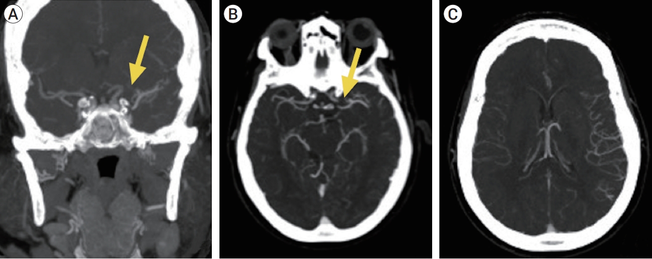
The NIHSS (National Institutes of Health Stroke Scale) score at admission was 4. Computed tomography (CT) angiography revealed a left M1 occlusion (Fig. 1). Because of the low NIHSS score and almost 7 hours after the ictus, no venous thrombolysis or interventional treatment was proposed. At that time, aspirin was prescribed.
Fifty-one hours after the TLKW, the patient had an NIHSS score of 11; a perfusion magnetic resonance imaging (MRI) revealed an ischemic core volume of 8 ml with a large mismatch ratio observed, and therefore, an ET was proposed (Fig. 2).
Groin puncture occurred 52 hours after the TLKW; subsequently a direct-aspiration first-pass technique (ADAPT) was used and one pass resulted in a thrombolysis in cerebral infarction scale (TICI) grade 3 recanalization.
The day after the procedure, an NIHSS score of 16 was observed; no hemorrhagic signals were detected by CT.
Five days after the procedure, the patient had an NIHSS score of 3 with a mild dysarthria and partial facial palsy. She was discharged to another hospital.
The patient is followed up on an outpatient basis. In the 6-month evaluation, the patient still presents with an NIHSS score of 2, being capable of self-care (Modified Rankin Scale 2).

DISCUSSION

This kind of case raises a huge discussion about whether patients can be treated outside of the standard therapeutic window and whether the type of collateral circulation can extend this window. It is already known that <30% [6] of patients with LVO have a “slow progressor” type and can benefit substantially from ET after 8 hours. This was demonstrated in the DAWN [11] and DEFUSE-3 [2] trials.
Our case presents that some patients might have a very slow progressor type, which we name “turtle” progressor type. These patients could possibly benefit from ET beyond the DAWN trial criteria (>24 hours) in highly selected cases. This progressor type might exist where there is an exceptional capacity to sustain collateral blood flow, and where the ischemic core does not become enlarged, protecting the penumbra area.
Another interesting point which is seen in this case is a major worsening the day after treatment (NIHSS score of 11 to 16) without hemorrhagic transformation despite successful endovascular treatment (TICI 3). In addition to the Contrast-induced encephalopathy already described by some authors [5], it could be that patients with the slow progressor type, and in our case the “turtle” progressor type, may suffer a cerebral hyperperfusion-like syndrome when a TICI grade 3 recanalization is achieved after LVO. Because recanalization changes the low-pressure cerebral flow in the microcirculation (collateral flow) to a high-pressure flow, a hyperperfusion-like syndrome that we call “collateral syndrome” could occur. This event needs to be evaluated in other patients with the collateral profile of slow progressor and “turtle” progressor to analyze the reproducibility of this kind of “collateral syndrome”.

CONCLUSIONS

This is a rare case demonstrating a very late thrombectomy 52 hours after the TLKW with a good recovery observed 5 days after the procedure.
In some cases, there appears to be a direct relationship between some types of collateral circulation (slow progressor and “turtle” progressor) and extension of the treatment window. Patients with these progressor types can experience a hyperperfusion-like syndrome (“collateral syndrome”) after the recanalization of an LVO, which can lead to clinical worsening in the 48 hours immediately after the ET. However, further studies are needed in order to evaluate the reproducibility of this hypothetical syndrome.

NOTES

Disclosure

The authors report no conflict of interest concerning the materials or methods used in this study or the findings specified in this paper.

Fig. 1.
Cranial CT angiography showing a left M1 occlusion (yellow arrow in A and B) with good collateral circulation to the left cerebral hemisphere (C). A-coronal view; B and C-axial view. CT, computed tomography.
jcen-2021-e2021-01-003f1.jpg
Fig. 2.
MRI and perfusion MRI at 51 hours after TLKW and endovascular thrombectomy 52 hours after TLKW. MRI before endovascular treatment DWI (A), FLAIR (B) and perfusion (C), Left ICA DSA image before mechanical thrombectomy Anteriorposterior (D), and after one pass of ADAPT (E). MRI, magnetic resonance imaging; TLKW, time last known well; DWI, diffusion-weighted imaging; FLAIR, fluid-attenuated inversion recovery; ICA, internal carotid artery; DSA, digital subtraction angiography; ADAPT, a direct-aspiration first-pass technique.
jcen-2021-e2021-01-003f2.jpg

REFERENCES

1. Aguilar-Salinas P, Santos R, Granja MF, Effendi S, Sauvageau E, Hanel R, et al. Revisiting the therapeutic time window dogma: successful thrombectomy 6 days after stroke onset. BMJ Case Reports. 2018 2018:bcr-2018-014039.
crossref pmid
2. Albers GW, Marks MP, Kemp S, Christensen S, Tsai JP, Ortega-Gutierrez S, et al. Thrombectomy for stroke at 6 to 16 hours with selection by perfusion imaging. N Engl J Med. 2018 Feb;378(8):708-18.
crossref pmid pmc
3. Berkhemer OA, Fransen PSS, Beumer D, van den Berg LA, Lingsma HF, Yoo AJ, et al. A randomized trial of intraarterial treatment for acute ischemic stroke. N Engl J Med. 2015 Jan;372(1):11-20.
pmid
4. Campbell BCV, Mitchell PJ, Kleinig TJ, Dewey HM, Churilov L, Yassi L, et al. Endovascular therapy for ischemic stroke with perfusion-imaging selection. N Engl J Med. 2015 Mar;372(11):1009-18.
crossref pmid
5. Chu YT, Lee KP, Chen CH, Sung PS, Lin YH, Lee CW, et al. Contrast-induced encephalopathy after endovascular thrombectomy for acute ischemic stroke. Stroke. 2020 Dec;51(21):3756-9.
crossref pmid
6. Copen WA, Rezai Gharai L, Barak ER, Schwamm LH, Wu O, Kamalian S, et al. Existence of the diffusion-perfusion mismatch within 24 hours after onset of acute stroke: Dependence on proximal arterial occlusion. Radiology. 2009 Mar;250(3):878-86.
crossref pmid
7. Desai SM, Haussen DC, Aghaebrahim A, Al-Bayati AR, Santos R, Nogueira RG, et al. Thrombectomy 24 hours after stroke: Beyond DAWN. J Neurointerv Surg. 2018 Nov;10(11):1039-42.
crossref pmid
8. Goyal M, Demchuk AM, Menon BK, Eesa M, Rempel JL, Thornton J, et al. Randomized assessment of rapid endovascular treatment of ischemic stroke. N Engl J Med. 2015 372:1019-30.
crossref pmid
9. Hacke W, Kaste M, Bluhmki E, Brozman M, Dávalos A, Guidetti D, et al. Thrombolysis with alteplase 3 to 4.5 hours after acute ischemic stroke. N Engl J Med. 2008 Sep;359(13):1317-29.
crossref pmid
10. Jovin TG, Chamorro A, Cobo E, Miquel MA, Molina CA, Rovira A, et al. Thrombectomy within 8 hours after symptom onset in ischemic stroke. N Engl J Med. 2015 372:2296-306.
crossref pmid
11. Nogueira RG, Jadhav AP, Haussen DC, Bonafe A, Budzik RF, Bhuva P, et al. Thrombectomy 6 to 24 hours after stroke with a mismatch between deficit and infarct. N Engl J Med. 2018 Jan;378(1):11-21.
pmid
12. Rocha M, Jovin TG. Fast versus slow progressors of infarct growth in large vessel occlusion stroke: clinical and research implications. Stroke. 2017 Sep;48(9):2621-7.
crossref pmid
13. Saver JL, Goyal M, Bonafe A, Diener HC, Levy EI. Pereiraet VM, et al. Stent-retriever thrombectomy after intravenous t-PA vs. t-PA alone in stroke. N Engl J Med. 2015 Jun;372(24):2285-95.
crossref pmid
14. So C, Chaudhry N, Gandhi D, Cole JW, Motta M. Endovascular thrombectomy in acute-onset ischemic stroke-beyond the standard time windows: A case report and a review of the literature. Case Rep Neurol. 2018 Sep;10(3):279-85.
crossref pmid pmc
TOOLS
METRICS Graph View
  • 2 Crossref
  •  0 Scopus
  • 3,076 View
  • 102 Download
ORCID iDs

Igor Pagiola
https://orcid.org/0000-0001-9052-7946

Related articles


ABOUT
BROWSE ARTICLES
EDITORIAL POLICY
FOR CONTRIBUTORS
Editorial Office
The Journal of Cerebrovascular and Endovascular Neurosurgery (JCEN), Department of Neurosurgery, Wonkwang University
School of Medicine and Hospital, 895, Muwang-ro, Iksan-si, Jeollabuk-do 54538, Korea
Tel: +82-2-2279-9560    Fax: +82-2-2279-9561    E-mail: editor.jcen@the-jcen.org                

Copyright © 2024 by Korean Society of Cerebrovascular Surgeons and Korean NeuroEndovascular Society.

Developed in M2PI

Close layer
prev next